Forecasting connectomics - a naive Kurzweilian approach
The connectome of an organism is a map of all neurons and their connections. This may be thought of as a graph with the neurons as nodes and synaptic connections as edges. However, to successfully simulate an organism’s brain using a connectome, more information will be needed. Here we take the term ‘connectome’ to refer to the graph and the underlying electron microscopy images of the neurons, which contain much more information.
The complete connectome of the nematode worm (Caenorhabditis Elegans) was published in 1986. A complete set of images of the fruit fly (Drosophila melanogaster) was published in 2018. However, all of the neurons and their connections have not yet been segmented or traced. In January 2020 researchers published the connectome of the central brain of the fruit fly, containing 25,000 neurons, which to my knowledge is the largest connectomics dataset published to date.
I thought it would be fun/interesting to plot the progress of connectomics over time and try to extrapolate out any trend observed. So, I did a literature search for all studies to date which either traced or segmented neurons and marked out synapses in electron microscopy data:
[1] D. D. Bock, et al. “Network anatomy and in vivo physiology of visual cortical neurons”, Nature 471 (7337) (2011) 177–182. doi:10.1038/nature09802.
[2] K. L. Briggman, M. Helmstaedter, W. Denk, Wiring specificity in the direction-selectivity circuit of the retina, Nature 471 (7337) (2011) 183–188.
[3] D. J. Bumbarger, M. Riebesell, C. Rodelsperger, R. J. Sommer, System-wide rewiring underlies behavioral differences in predatory and bacterial-feeding nematodes, Cell 152 (1-2) (2013) 109–119.
[4] C.-Y. Lin, et al., A comprehensive wiring diagram of the protocerebral bridge for visual information processing in the drosophila brain, Cell Reports 3 (5) (2013) 1739–1753. [link]
[5] S. ya Takemura, et al., A visual motion detection circuit suggested by drosophila connectomics, Nature 500 (7461) (2013) 175–181. [link]
[6] M. Helmstaedter, K. L. Briggman, S. C. Turaga, V. Jain, H. S. Seung, W. Denk, Connectomic reconstruction of the inner plexiform layer in the mouse retina, Nature 500 (7461) (2013) 168–174. [link]
[7] N. Kasthuri, et al., Saturated reconstruction of a volume of neocortex, Cell 162 (3) (2015) 648–661. [link]
[8] A. A. Wanner et al., 3-dimensional electron microscopic imaging of the zebrafish olfactory bulb and dense reconstruction of neurons, Scientific Data 3 (1). [link]
[9] K. Ryan, Z. Lu, I. A. Meinertzhagen, The CNS connectome of a tadpole larva of Ciona intestinalis (l.) highlights sidedness in the brain of a chordate sibling, eLife 5 (2016) [link]
[10] S.-y. Takemura, et al., A connectome of a learning and memory center in the adult Drosophila brain, eLife 6 (2017). [link]
[11] K. Eichler, et al., The complete connectome of a learning and memory centre in an insect brain, Nature 548 (7666) (2017) 175–182. [link]
[12] C. S. Xu, et al., A connectome of the adult drosophila central brain (preprint) [link]
[13] L. K. Scheffer, et al., A connectome and analysis of the adult drosophila central brain, eLife 9 (2020). [link]
[14] J. S. Phelps, et al., Reconstruction of motor control circuits in adult drosophila using automated transmission electron microscopy, Cell 184 (3) (2021) 759–774.e18. [link]
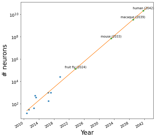
Next I plotted most of the # neurons data vs the date of publication:
Next I did linear regression on the (year, log(# neurons)) data which is equivalent to fitting an exponential function to the data. (The reason for fitting the data in this way was to avoid the bias that occurs when fitting an exponential function with least squares regression that leads to the y larger values being fit more accurately than smaller ones.) After doing the linear regression I extrapolated it forward in time.
The projection for the fruit fly connectome (2024) seems about right. If anything, we may see it slightly sooner. It will be interesting to see how much longer it will take before we have physically realistic models of the fruit fly and fruit fly behavior, something my friend Logan T. Collins has advocated for. A project to produce the mouse brain connectome is underway, and again, the date extrapolated to — 2033 — seems plausible. Beyond that though, I have very little idea how plausible the projections are!
Here’s some numbers that show the challenges just with scanning the entire brain (not to mention segmenting/tracing all the neurons accurately!):
Assuming an isotropic voxel size of 20 nm, it is estimated that storing the images of an entire human brain would require 175 exabytes of storage. It seems we are approaching hard drives which cost about 1.5 cents per gigabyte. Even at those exorbitantly low prices, it would still cost $2.6 billion to store all those images!
The volume of the human brain is about 1.2x10^6 cubic millimeters. The Zeiss MultiSEM contains either 61 or even 91 electron beams which scan a sample in parallel. According to a Zeiss video presentation from April 8th, 2020, it can scan a 1x1mm area at 4nm resolution in 6.5 minutes. Assuming a slice thickness of 20 nn, a single such machine would require 742,009 years to scan the entire brain!
X-ray holographic nano-tomography might be the path forward …


Zeiss will make 20k beam SEM which will suffice to get whole mouse connectome done in under a year.
This might be a dumb question but what do you do once you’ve mapped out these edges and nodes?